Skip to content
Last Updated on April 12, 2025 by John Hookway
Right now, someone is losing their sight. And the tragedy? It didn’t have to happen.
Blindness feels like a random act of cruel fate—until you learn that 80% of vision loss worldwide could be stopped before it starts. This isn’t wishful thinking—it’s medical fact.
Think about what you’ve seen today: a child’s smile, text messages from friends, the colors of your morning coffee mug. Now imagine those memories becoming your last visual experiences.
For 43 million people globally living with blindness, this isn’t hypothetical. But here’s what most don’t know: the path to darkness often has warning signs we miss or ignore.
“I just thought I needed stronger glasses,” says Maria, 62, diagnosed with advanced glaucoma after years of dismissing her gradual vision changes. By then, her permanent vision loss could have been prevented with early treatment.
The human eye contains 107 million light-sensitive cells, yet we take this miracle for granted. We protect our homes with security systems and our cars with insurance—but what about the organs responsible for 80% of how we experience the world?
What if five minutes of reading could save your sight? What if simple habits could protect your family’s vision for decades? What if the most devastating eye conditions announced themselves early enough to stop?
The answers might surprise you. They might also save your sight.
Let’s explore the facts about eye blindness everyone should know—because vision preservation isn’t about luck. It’s about knowledge, action, and understanding that what you don’t know absolutely can hurt you.
What is Eye Blindness?

Eye blindness describes a condition where a person experiences significant vision loss that cannot be corrected with standard methods like glasses, contact lenses, or medication.
This condition affects millions worldwide and can severely impact daily activities such as reading, driving, recognizing faces, and navigating environments.
Vision impairment exists on a spectrum, from mild to severe, with blindness representing the most severe form.
Example(s) of Eye Blindness
Legal blindness and total blindness represent two distinct categories of vision impairment, each with specific characteristics and implications for those affected.
Legal blindness is defined as having a visual acuity of 20/200 or worse in the better-seeing eye, even with correction. This means a person with legal blindness must stand 20 feet away to see what someone with normal vision can see from 200 feet away.
Alternatively, legal blindness can be diagnosed when a person’s visual field is restricted to 20 degrees or less (tunnel vision), compared to the normal visual field of approximately 180 degrees.
People with legal blindness may still have some useful vision and can often perceive light, colors, and shapes to varying degrees.
Total blindness, in contrast, refers to the complete absence of visual perception. Individuals with total blindness cannot perceive light, distinguish shapes, or detect movement. This condition represents about 15% of the visually impaired population worldwide.
Despite common misconceptions, many people who identify as blind have some degree of light perception or residual vision.
According to the World Health Organization, approximately 43 million people globally live with blindness, while 295 million experience moderate to severe visual impairment.
Types of Eye Blindness
Eye blindness generally falls into two main categories based on when and how the condition develops: congenital blindness and acquired blindness. Each type has distinct causes, presentations, and treatment approaches.
Congenital Blindness
Congenital blindness refers to vision loss that is present at birth or develops during the first few months of life. This condition affects approximately 1.4 million children worldwide, with higher rates in developing countries.
Genetic disorders account for a significant portion of congenital blindness cases. Conditions such as Leber congenital amaurosis, retinitis pigmentosa, and albinism result from genetic mutations that affect eye development or function. These conditions are often inherited in various patterns—autosomal dominant, autosomal recessive, or X-linked—and may appear in family histories.
Birth complications also contribute to congenital blindness. Premature birth increases the risk of retinopathy of prematurity, a condition where abnormal blood vessel growth damages the retina. Maternal infections during pregnancy, particularly those causing TORCH syndrome (Toxoplasmosis, Other infections, Rubella, Cytomegalovirus, and Herpes simplex), can severely impact fetal eye development.
An interesting fact about people born blind is that they experience dreams differently than sighted individuals. Rather than visual imagery, their dreams are composed of sounds, tactile sensations, emotions, and smells. This demonstrates how the brain adapts to process and interpret information based on available sensory inputs.
Acquired Blindness
Acquired blindness develops after birth, often later in life, and can result from various factors including disease progression, injuries, or the natural aging process.
Disease-related blindness accounts for most acquired vision loss cases. Cataracts remain the leading cause worldwide, affecting over 65 million people and accounting for 51% of global blindness.
Glaucoma, the second most common cause, damages the optic nerve through increased eye pressure. Diabetic retinopathy affects approximately one-third of people with diabetes and can lead to severe vision loss if left untreated.
Age-related macular degeneration (AMD) primarily affects older adults, causing the breakdown of the macula—the central portion of the retina responsible for sharp, central vision. AMD affects more than 196 million people globally.
Trauma and injuries represent another significant cause of acquired blindness. Workplace accidents, sports injuries, chemical burns, and combat-related trauma can cause immediate or progressive vision loss. According to occupational safety statistics, eye injuries account for approximately 2,000 workplace injuries daily in the United States alone.
“To shut your eyes is to guess nothing of blindness. Beneath your world of skies and faces and buildings exists a rawer and older world, a place where surface planes disintegrate and sounds ribbon in shoals through the air,” wrote Anthony Doerr in “All the Light We Cannot See,” capturing how blindness creates a fundamentally different sensory experience.
The duration of blindness varies significantly based on its cause. Temporary blindness might result from conditions like migraines, concussions, or certain medications, typically resolving within hours or days.
Progressive conditions like glaucoma or retinitis pigmentosa may cause gradual vision loss over years or decades. Sudden blindness from events like retinal detachment or stroke requires immediate medical attention, as prompt treatment within hours can sometimes restore vision.
Permanent blindness results when the visual pathway—from the eye to the brain’s visual processing centers—sustains irreversible damage.
Color blindness presents a related but distinct vision condition affecting about 8% of men and 0.5% of women worldwide. Most color-blind individuals aren’t truly “blind” to colors but instead have difficulty distinguishing between certain color combinations.
The most common form, red-green color blindness, makes it challenging to differentiate these two colors and their combinations.
Interestingly, some color-blind individuals develop enhanced abilities to detect camouflage patterns and subtle variations in texture, which historically provided evolutionary advantages for hunting. C
urrent research explores gene therapy approaches that have successfully restored color vision in some animal models and shown promising results in early human trials.

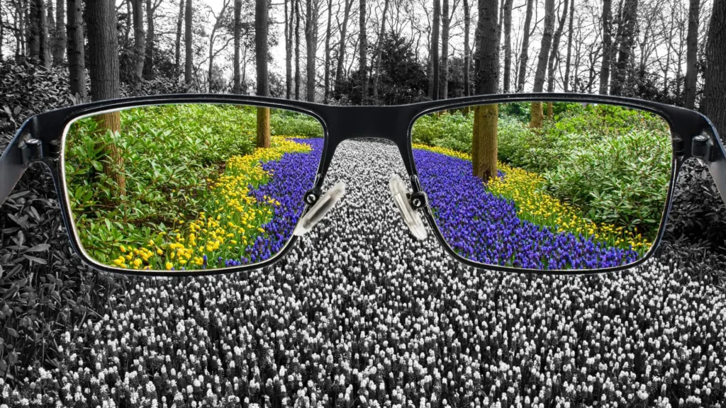
As shown in the pie chart:
- 80% of blindness is acquired, often from conditions like cataracts, glaucoma, and diabetes.
- Only 20% is congenital, linked to genetics or complications during pregnancy or birth.
Key WHO Insight on Preventable Blindness
“At least 1 billion people have a vision impairment that could have been prevented or has yet to be addressed.”
— World Health Organization
Benefits of Preventative Eye Care
Regular eye exams catch diseases before symptoms appear, preventing 95% of severe vision loss cases. Proper eye protection and nutrition reduce risk factors by up to 60%. Early intervention can halt or reverse progression of most common eye conditions
Regular Eye Exams
Regular eye examinations form the foundation of preventative eye care. These exams go far beyond simply checking if you need glasses.
They serve as the first line of defense against potentially sight-threatening conditions that develop silently.
According to research published in the British Journal of Ophthalmology, nearly 75% of cases of permanent vision loss were preventable or treatable if detected early enough through regular screenings.
During a comprehensive eye exam, ophthalmologists can detect the earliest signs of conditions like glaucoma, diabetic retinopathy, and macular degeneration—often years before a patient notices any symptoms.
This early detection window is critical because most eye diseases progress gradually and painlessly. By the time vision changes become noticeable to the patient, significant and sometimes irreversible damage may have already occurred.
“A comprehensive dilated eye exam by an eye doctor can find eye diseases in the early stages. This is when treatment to prevent vision loss is most effective,” notes the National Eye Institute.
The American Academy of Ophthalmology recommends adults get a baseline eye examination at age 40, the time when early signs of disease and vision changes may start to occur.
After age 60, examinations should occur annually, as the risk for eye disease increases dramatically with age.
Early Detection Benefits
The benefits of early detection through regular exams cannot be overstated. For conditions like glaucoma, which damages the optic nerve, early intervention can slow progression by up to 90% with appropriate treatment.
Once vision is lost to glaucoma, it cannot be restored, making early detection the only defense against blindness from this condition.
For diabetic retinopathy, the leading cause of blindness in working-age adults, studies show that early detection and treatment reduce the risk of blindness by 95%.
The Diabetes Control and Complications Trial demonstrated that tight blood sugar control—often prompted by early detection of retinopathy during an eye exam—reduced the progression of diabetic eye disease by 76%.
The cost-benefit analysis of regular eye exams is clear. A study in the Journal of Health Economics found that the lifetime cost of blindness far exceeds the cumulative cost of regular eye examinations and early intervention.
For every dollar spent on preventative eye care, approximately $5 is saved in treatment costs and productivity losses from advanced eye disease.
Eye Health Practices
Beyond regular examinations, daily eye health practices significantly reduce the risk of vision loss. These practices act as a frontline defense against environmental and lifestyle factors that contribute to eye damage over time.
UV protection stands as one of the most critical yet often overlooked aspects of eye health. Studies from the World Health Organization indicate that up to 20% of cataracts may be caused by UV exposure.
“UV radiation, whether from natural sunlight or indoor artificial rays, can damage the eye’s surface tissues as well as the cornea and lens. By wearing UV-blocking sunglasses, you can enjoy the summer safely while lowering your risk for potentially blinding eye diseases and tumors,” explains Dr. Michael Kutryb, MD, from the American Academy of Ophthalmology.
The type of protection matters significantly. Research published in the journal Ophthalmology demonstrated that wraparound sunglasses that block 99-100% of UVA and UVB radiation provide the most effective protection.
Even on cloudy days, UV radiation penetrates atmospheric cover, making year-round protection essential.
Digital eye strain represents another modern challenge to eye health. The average American spends over 7 hours daily on digital devices, leading to symptoms collectively known as Computer Vision Syndrome.
Implementing the 20-20-20 rule (looking at something 20 feet away for 20 seconds every 20 minutes) reduces eye strain by giving eye muscles a chance to relax. Studies show this simple practice can decrease symptoms of digital eye strain by up to 60%.
Nutritional Support for Vision
Nutrition plays a fundamental role in maintaining eye health and preventing vision loss. The Rotterdam Study, which followed over 4,000 participants, found that those with high dietary intake of vitamins C and E, beta-carotene, and zinc had a 35% lower risk of developing age-related macular degeneration compared to those with low intake.
Specific nutrients have been identified as particularly beneficial for eye health:
- Omega-3 fatty acids found in fish reduce inflammation and help prevent dry eye syndrome
- Lutein and zeaxanthin, found in leafy greens, act as natural sunglasses, absorbing harmful blue light
- Vitamin A, present in orange vegetables, is essential for the proper functioning of the retina
- Vitamin C helps maintain the health of blood vessels in the eye
The landmark Age-Related Eye Disease Study (AREDS) demonstrated that a specific formulation of vitamins C and E, beta-carotene, zinc, and copper reduced the risk of advanced AMD by about 25% over a six-year period.
The follow-up AREDS2 study refined this formulation by adding lutein and zeaxanthin while removing beta-carotene, making it safer for smokers while maintaining efficacy.
For those seeking to improve their nutritional support for eye health, the book “Feed Your Eyes Right” by Dr. Laurie Capogna provides detailed dietary recommendations specifically tailored to prevent common eye diseases.
Lifestyle Modifications
Lifestyle factors significantly impact long-term eye health and can either accelerate or slow the progression of many eye conditions.
Research consistently shows that smoking multiplies the risk of cataracts by 2-3 times and increases the risk of AMD by 2-4 times. The British Journal of Ophthalmology reports that smoking causes oxidative stress, reduces blood flow to the eyes, and depletes antioxidant levels—all factors that damage eye tissues.
Physical activity, often overlooked in eye health discussions, plays a critical role in preventing vision loss. A 2020 study in the American Journal of Ophthalmology found that regular exercise reduced the risk of glaucoma by up to 25% by improving blood flow to the optic nerve and reducing intraocular pressure. Even moderate activity like brisk walking for 30 minutes daily provides significant protective benefits.
Blood pressure and diabetes management directly correlate with eye health. Hypertension can damage the delicate blood vessels in the retina, while uncontrolled diabetes accelerates the development of diabetic retinopathy.
The Beaver Dam Eye Study demonstrated that for every 10 mmHg increase in systolic blood pressure, the risk of retinopathy increased by 10-30%, depending on other risk factors.
Workplace Eye Safety
Occupational eye injuries account for over 2,000 cases of preventable blindness annually in the United States alone.
These injuries disproportionately affect manufacturing, construction, and healthcare workers. Proper eye protection prevents 90% of these injuries, according to the American Academy of Ophthalmology.
Safety standards for eye protection vary by industry, but ANSI Z87.1-certified eyewear provides a baseline for impact protection.
For workers exposed to additional hazards like chemicals, radiation, or extreme temperatures, specialized protection is essential. The National Institute for Occupational Safety and Health offers industry-specific guidelines that, when followed, dramatically reduce injury rates.
Beyond acute injuries, chronic workplace conditions can also threaten eye health. Poor lighting increases eye strain and fatigue, while excessive glare from screens or reflective surfaces damages retinal cells over time.
Ergonomic adjustments such as proper monitor positioning (20-30 inches from the eyes, slightly below eye level) reduce strain by 35% according to workplace health studies.
Organizations implementing comprehensive eye safety programs report up to an 85% reduction in eye-related incidents and associated costs.
These programs typically combine proper equipment, regular training, and environmental modifications to create a culture of eye safety awareness.
Genetic Risk Assessment
Emerging research in genetic risk assessment offers a new frontier in preventative eye care. Many eye conditions have hereditary components, with family history increasing risk by 2-10 times depending on the condition and degree of relationship.
Genetic testing can now identify specific markers associated with conditions like glaucoma, AMD, and retinitis pigmentosa.
For those with identified genetic risk factors, proactive monitoring and intervention strategies can be implemented years before clinical symptoms might appear.
The field of pharmacogenomics is exploring how genetic profiles influence response to eye medications, potentially allowing for personalized treatment approaches that maximize effectiveness while minimizing side effects.
Dr. Edwin Stone’s book “The Genetics of Eye Disease” provides an accessible overview of how genetic factors influence eye health and how this knowledge can be applied to prevention strategies.
While genetic testing has limitations and ethical considerations, it represents a powerful tool for those with family histories of eye disease to take control of their eye health destiny.
When combined with traditional preventative measures, genetic risk assessment creates a comprehensive approach to eye care that addresses both inherited and environmental risk factors. This multi-faceted strategy represents the future of blindness prevention, where each individual receives care tailored to their unique risk profile.

Common Causes of Blindness
Major preventable causes: cataracts, glaucoma, and macular degeneration affect millions globally. Early detection and treatment can prevent up to 80% of blindness cases. Regular eye exams are critical for high-risk groups including older adults and diabetics
Globally, at least 2.2 billion people have vision impairment or blindness, with at least 1 billion cases being preventable or untreated.
This staggering number reflects conditions that, with proper care and early intervention, might never progress to severe vision loss. Understanding these common causes is the first step toward prevention.
Cataracts
Cataracts develop when proteins in the eye’s lens break down and clump together, creating a cloudy area that blocks light from passing through clearly.
This clouding process typically happens slowly over years, often starting after age 40, though many people don’t notice symptoms until their 60s.
Cataracts affect approximately 20.5 million Americans aged 40 and older, and by age 80, more than half of all Americans develop cataracts.
Globally, they account for 51% of worldwide blindness, particularly in regions with limited surgical services. The condition manifests as increasingly blurred vision, faded colors, glare sensitivity, and difficulty with night vision.
What makes cataracts particularly significant in discussions about preventable blindness is that surgical treatment is both widely available and highly effective.
The procedure involves removing the clouded lens and replacing it with an artificial one called an intraocular lens (IOL). Most patients experience dramatic improvement in vision almost immediately after surgery, with success rates above 95%.
Several factors increase cataract risk, including:
- Aging (primary factor)
- Extended UV light exposure without protection
- Smoking
- Diabetes
- Previous eye injury or inflammation
- Family history
- Long-term steroid medication use
Preventive strategies focus on limiting these risk factors. Wearing quality sunglasses that block 99-100% of UVA and UVB radiation, stopping smoking, controlling diabetes, and maintaining regular eye examinations can significantly reduce risk or catch cataracts early.
Glaucoma
Glaucoma represents a group of eye conditions that damage the optic nerve, often due to abnormally high pressure within the eye.
This pressure, called intraocular pressure (IOP), builds when the fluid that normally flows through the eye cannot drain properly. The damage occurs gradually and can lead to permanent vision loss if untreated.
Glaucoma affects about 2.2 million Americans aged 40 and older, with another 2 million unaware they have the condition.
This “silent thief of sight” typically causes no symptoms in its early stages, which makes regular screening essential. By the time vision changes become noticeable, permanent damage has often occurred.
Types of Glaucoma and Their Mechanisms
Open-angle glaucoma, the most common form, develops slowly when the drainage canals become clogged over time.
Angle-closure glaucoma occurs when the iris blocks the drainage angle, causing a sudden rise in eye pressure – a medical emergency requiring immediate treatment.
Normal-tension glaucoma presents a particular challenge, as it damages the optic nerve even with normal IOP levels.
This variant appears associated with blood flow abnormalities and may have genetic links. Recent research suggests that vascular factors, including blood pressure fluctuations and nutrient delivery to the optic nerve, play significant roles.
Treatment focuses primarily on lowering intraocular pressure through:
- Prescription eye drops that either reduce fluid production or improve drainage
- Oral medications (typically carbonic anhydrase inhibitors)
- Laser treatments that improve drainage system function
- Minimally invasive glaucoma surgeries (MIGS)
- Traditional surgeries like trabeculectomy for advanced cases
Dr. Robert Weinreb’s research at the Hamilton Glaucoma Center suggests that beyond pressure management, neuroprotection strategies may soon become important treatment components. These approaches aim to protect nerve cells from damage even when pressure control is suboptimal.
Progression monitoring involves sophisticated visual field testing and optical coherence tomography to track minute changes in optic nerve structure and function. This allows physicians to adjust treatment before significant vision loss occurs.
Macular Degeneration
Age-related macular degeneration (AMD) progressively destroys sharp central vision by affecting the macula – the central portion of the retina responsible for fine-detail vision.
This condition primarily impacts adults over 50 and is the leading cause of blindness among older adults in developed countries.
AMD exists in two main forms:
- Dry AMD (85-90% of cases): Characterized by thinning of the macula and accumulation of small yellow deposits called drusen under the retina. It typically progresses slowly over years.
- Wet AMD (10-15% of cases): Involves abnormal blood vessel growth under the retina that can leak fluid and blood. This form progresses much more rapidly and can cause severe vision loss within months if untreated.
The precise cause remains incompletely understood, but several risk factors have been identified:
- Age (primary factor)
- Smoking (doubles risk)
- Family history and genetic factors
- High blood pressure and cardiovascular disease
- Obesity
- Diet low in antioxidants and omega-3 fatty acids
- Prolonged sun exposure
Research from the Age-Related Eye Disease Studies (AREDS and AREDS2) demonstrated that specific nutritional supplements can slow progression in intermediate to advanced dry AMD. These supplements typically contain vitamins C and E, zinc, copper, lutein and zeaxanthin.
For wet AMD, treatment advances have been remarkable. Anti-VEGF (vascular endothelial growth factor) injections like ranibizumab, aflibercept, and bevacizumab have revolutionized care by blocking the growth of abnormal blood vessels.
These medications, administered directly into the eye, have transformed wet AMD from a rapidly blinding condition to a manageable chronic disease for many patients.
In his book “Macular Degeneration: The Complete Guide to Saving and Maximizing Your Sight,” ophthalmologist Lylas G. Mogk provides excellent guidance for patients on living well with AMD, including practical strategies for vision rehabilitation and home modifications.
Diabetic Retinopathy
Diabetic retinopathy occurs when chronically high blood sugar damages the tiny blood vessels in the retina.
It is the leading cause of blindness among American adults, affecting 4.1 million people in the U.S., with nearly 900,000 at risk of severe vision loss.
The condition progresses through several stages if left untreated:
- Mild nonproliferative retinopathy: Small areas of balloon-like swelling in retinal blood vessels
- Moderate nonproliferative retinopathy: Blood vessels that nourish the retina become blocked
- Severe nonproliferative retinopathy: More blood vessels become blocked, depriving areas of the retina of blood supply
- Proliferative retinopathy: New, abnormal blood vessels grow on the retina’s surface that can rupture and bleed
What makes diabetic retinopathy particularly tragic is its high rate of preventability. Tight blood glucose control significantly reduces both development and progression risk.
The landmark Diabetes Control and Complications Trial showed that intensive diabetes management reduced retinopathy risk by 76% in people without existing eye damage.
Regular comprehensive dilated eye exams can catch changes before symptoms develop. Treatment options include:
- Laser therapy to seal leaking blood vessels
- Anti-VEGF injections to reduce abnormal vessel growth
- Vitrectomy surgery for advanced cases with significant bleeding
Dr. Lloyd Paul Aiello from Harvard’s Joslin Diabetes Center has pioneered research into the biochemical pathways of diabetic retinopathy, potentially leading to new drug targets beyond current treatments.
Refractive Errors and Corneal Diseases
Uncorrected refractive errors—myopia (nearsightedness), hyperopia (farsightedness), astigmatism, and presbyopia—represent the most common vision problems worldwide.
While not typically considered causes of blindness in developed countries where glasses and contact lenses are readily available, they remain leading causes of functional blindness in developing regions.
The global myopia epidemic deserves special attention. Rates have skyrocketed, particularly in East Asian countries where up to 90% of young adults are myopic.
High myopia increases risks for serious eye conditions including retinal detachment, glaucoma, and myopic macular degeneration. Environmental factors, particularly reduced outdoor time during childhood and increased near-work activities, appear to play significant roles in this trend.
Corneal diseases represent another important category of potentially blinding conditions. Keratoconus, a progressive thinning of the cornea that causes it to bulge outward, affects approximately 1 in 2,000 people.
Early detection allows for interventions like corneal crosslinking, which can halt progression. Corneal transplantation remains the definitive treatment for advanced cases.
Infectious keratitis (corneal infection) presents another preventable cause of blindness, particularly in contact lens wearers.
Proper lens hygiene, avoiding sleeping in contacts, and prompt attention to eye redness or pain can prevent these infections from causing permanent damage.
For those interested in exploring corneal diseases further, “Cornea” by Drs. Krachmer, Mannis, and Holland serves as the definitive reference text on the subject, offering detailed information on diagnosis and management strategies.

How does Eye Care Work?
Eye care represents a comprehensive approach to maintaining vision health and preventing vision loss. Unlike reactive medical care that addresses problems after they occur, eye care focuses on proactive measures and regular monitoring to identify problems before they become serious.
This proactive approach explains why most blindness cases can be prevented through proper care routines.
Step #1: Regular Check-ups
The foundation of effective eye care is consistent professional monitoring. Regular eye exams serve as an early warning system for potential vision problems, allowing for intervention before permanent damage occurs.
For adults with no risk factors, eye specialists typically recommend comprehensive eye exams every 1-2 years. However, people with diabetes, a family history of eye disease, or those over 60 should schedule exams more frequently—often annually or as recommended by their eye care professional.
During these exams, eye doctors perform several tests to assess your visual acuity, eye pressure, peripheral vision, and the health of your retina and optic nerve.
A comprehensive eye exam includes several key components that work together to provide a complete picture of your eye health:
- Visual acuity testing using standard eye charts to measure how well you see at various distances
- Refraction assessment to determine if you need corrective lenses
- Eye pressure testing to screen for glaucoma
- Examination of the front part of the eye using a special microscope called a slit lamp
- Retinal examination to check the back of your eye for signs of disease
Finding the Right Eye Care Provider
Working with the right eye care professional is essential for effective prevention. There are three main types of eye care providers:
- Ophthalmologists: Medical doctors who specialize in eye care, provide comprehensive eye care including surgery
- Optometrists: Eye doctors who provide primary vision care, including vision tests and corrective lenses
- Opticians: Technicians who fit and adjust eyeglasses and contact lenses
For routine eye care, an optometrist can handle most needs. However, if you have a family history of eye disease or other risk factors, an ophthalmologist may be more appropriate.
When selecting a provider, check their credentials, experience with your specific concerns, and whether they accept your insurance.
Step #2: Lifestyle Adjustments
Daily habits significantly impact long-term eye health. Making consistent lifestyle adjustments helps protect your vision from cumulative damage that can lead to blindness.
One of the most important adjustments is protecting your eyes from harmful light sources. Ultraviolet (UV) radiation from the sun can damage the eyes over time, contributing to conditions like cataracts and macular degeneration.
Blue light from digital screens can cause digital eye strain and may disrupt sleep patterns when exposure occurs close to bedtime.
To protect your eyes from harmful light:
- Wear sunglasses that block 99-100% of UVA and UVB radiation whenever outdoors
- Use blue light filters on digital devices or wear computer glasses with blue light filtering technology
- Follow the 20-20-20 rule when working with screens: every 20 minutes, look at something 20 feet away for 20 seconds
- Ensure proper lighting in your workspace to reduce eye strain
- Position your computer screen slightly below eye level and about arm’s length away
Nutritional Support for Eye Health
Diet plays a crucial role in maintaining eye health. Specific nutrients help protect the structures of the eye and support visual function.
Research shows that maintaining adequate levels of certain nutrients can reduce the risk of developing serious eye conditions.
Key nutrients for eye health include:
- Omega-3 fatty acids: Found in fatty fish like salmon and tuna, these reduce inflammation and may help prevent dry eyes
- Lutein and zeaxanthin: Present in leafy greens like spinach and kale, these antioxidants filter harmful blue light
- Vitamins C and E: Found in citrus fruits and nuts respectively, these antioxidants protect cells from damage
- Zinc: Present in beans, nuts, and whole grains, zinc helps vitamin A reach the retina
- Vitamin A: Found in orange vegetables like carrots and sweet potatoes, essential for night vision

Step #3: Manage Chronic Conditions
Certain chronic medical conditions significantly increase the risk of developing vision problems. Managing these conditions effectively is a critical part of preventative eye care.
Diabetes presents one of the greatest risks to vision health. High blood sugar damages the small blood vessels in the retina, leading to diabetic retinopathy—a leading cause of blindness. People with diabetes should:
- Maintain tight control of blood sugar levels as recommended by their doctor
- Monitor blood pressure and cholesterol levels regularly
- Schedule comprehensive dilated eye exams at least annually
- Report any vision changes to their eye doctor immediately
- Work with their healthcare team to establish a diabetes management plan
Hypertension (high blood pressure) can also damage blood vessels in the eyes, potentially leading to bleeding, blurred vision, and even blindness. Managing blood pressure through medication, diet, exercise, and stress reduction helps protect your eyes.
Occupational Eye Protection
Many jobs carry risks for eye injuries or strain. Taking proper precautions in work environments can prevent serious vision damage.
For those working with hazardous materials or in environments with airborne particles:
- Wear appropriate safety eyewear certified by ANSI (American National Standards Institute)
- Ensure safety glasses have side shields when working with chemicals
- Use face shields or goggles for additional protection in high-risk environments
- Keep emergency eyewash stations accessible and know how to use them
- Regularly inspect and replace damaged protective eyewear
For office workers and others who use computers extensively:
- Position monitors at arm’s length and slightly below eye level
- Use matte screen filters to reduce glare
- Ensure proper lighting that doesn’t create glare on screens
- Take regular breaks using the 20-20-20 rule
- Consider computer glasses with slight magnification if you’re over 40
Step #4: Address Vision Changes Promptly
Early intervention significantly improves outcomes for most eye conditions. Learning to recognize warning signs and seeking prompt care can prevent permanent vision loss.
Common warning signs that require immediate attention include:
- Sudden blurry vision or difficulty focusing
- Floaters (small dark spots or strings that drift across your vision) or flashes of light
- Loss of peripheral vision
- Pain in or around the eye
- Redness or swelling of the eye
- Persistent dryness or tearing
If you experience any of these symptoms, contact your eye care professional promptly. Some conditions, like retinal detachment or acute angle-closure glaucoma, require emergency treatment to prevent permanent vision loss.
Creating an Eye Emergency Plan
Having a plan in place for eye emergencies can save critical time when vision is at risk. Your eye emergency plan should include:
- Your eye doctor’s emergency contact information
- Location of the nearest eye emergency service
- A list of your current medications and any known allergies
- Information about your vision insurance
- A designated person who can drive you to appointments if needed
7 Tips/Strategies for Preventative Eye Care
Tip #1: Wear Sunglasses
Quality sunglasses do more than make you look good—they provide essential protection against UV radiation that can damage your eyes over time.
When selecting sunglasses, look for pairs that block 99-100% of both UVA and UVB radiation. Wraparound styles provide better protection by blocking light from entering from the sides. Polarized lenses reduce glare, which is particularly helpful when driving or near water.
Wearing sunglasses consistently—even on cloudy days when UV rays still penetrate—reduces your risk of developing cataracts and macular degeneration. For added protection, wear a wide-brimmed hat along with your sunglasses when outdoors for extended periods.
Tip #2: Eat Essential Nutrients
Diet plays a key role in eye health maintenance. Research supports that certain nutrients can protect against age-related eye conditions.
The Age-Related Eye Disease Study (AREDS) found that a specific formula of vitamins C and E, beta-carotene, zinc, and copper can slow the progression of age-related macular degeneration. The follow-up study, AREDS2, found that adding lutein and zeaxanthin was also beneficial.
To support eye health through diet:
- Eat fatty fish like salmon or tuna twice a week for omega-3 fatty acids
- Include dark, leafy greens such as spinach, kale, and collard greens in your meals daily
- Snack on nuts, especially walnuts and almonds, for vitamin E
- Add citrus fruits and berries to your diet for vitamin C
- Include eggs in your diet for lutein, zeaxanthin, and zinc
Tip #3: Practice the 20-20-20 Rule
Digital eye strain has become increasingly common as screen use has grown. The 20-20-20 rule helps reduce this strain during extended screen time.
Every 20 minutes, take a 20-second break to look at something 20 feet away. This simple practice helps reset your focus and reduces eye fatigue. Set a timer on your phone or computer as a reminder until this becomes a habit.
Beyond the 20-20-20 rule, try to take longer breaks—about 5-10 minutes—every hour of screen use. During these breaks, stand up, stretch, and move around to reduce overall physical tension that can contribute to eye strain.
Tip #4: Maintain Proper Humidity
Dry eyes can result from environmental factors like low humidity, air conditioning, or heating systems. Creating the right environment helps protect your eyes from dryness and irritation.
Use a humidifier in your home and office to maintain optimal humidity levels (around 45-55%). Position computer screens to avoid air from fans, heating, or air conditioning blowing directly toward your eyes. When outdoors in windy or dry conditions, wear wraparound sunglasses to protect your eyes from drying elements.
If you experience persistent dry eyes despite these measures, speak with your eye doctor about artificial tears or other treatments that might help maintain proper eye lubrication.
Tip #5: Quit Smoking
Smoking damages nearly every part of the body, including the eyes. Research consistently shows that smokers have a significantly higher risk of developing age-related macular degeneration, cataracts, and optic nerve damage.
The toxic compounds in cigarette smoke cause oxidative stress and inflammation that damage eye tissues. They also reduce blood flow to the eyes and deplete antioxidant levels that protect eye cells.
If you smoke, quitting is one of the most important steps you can take for your eye health. Talk to your doctor about smoking cessation programs, medications, and support groups that can help you quit successfully.
Tip #6: Control Blood Sugar and Blood Pressure
Systemic health conditions directly impact eye health. Managing chronic conditions helps protect your vision over the long term.
High blood sugar damages the tiny blood vessels in the retina, leading to diabetic retinopathy. Similarly, hypertension can cause retinopathy, choroidopathy (affecting the layer of blood vessels beneath the retina), and optic neuropathy (damage to the optic nerve).
Work with your healthcare provider to:
- Monitor your blood sugar levels regularly if you have diabetes
- Check your blood pressure frequently if you have hypertension
- Take prescribed medications consistently
- Follow recommended dietary guidelines for your condition
- Exercise regularly as approved by your doctor
- Attend all scheduled check-ups for both your primary condition and eye health
Tip #7: Stay Physically Active
Regular physical activity benefits eye health in several ways. Exercise improves circulation, including blood flow to the eyes, and helps control conditions that can damage vision.
Research suggests that regular exercise may reduce the risk of developing glaucoma by lowering intraocular pressure. It also helps control blood sugar levels and blood pressure, reducing the risk of diabetic retinopathy and hypertensive eye disease.
Aim for at least 150 minutes of moderate aerobic activity per week, as recommended by health guidelines. Activities like walking, swimming, cycling, and dancing all provide benefits. Before starting any new exercise program, especially if you have existing eye conditions, consult with your healthcare provider.

Eye Health Myths Debunked
Understanding what truly works for eye health can make the difference between keeping your vision healthy and wasting time on ineffective practices. After learning how eye care works systematically, it’s important to clear up common misconceptions that might lead you astray.
Myth #1: Eating Carrots Improves Vision
The belief that carrots dramatically improve eyesight has persisted for decades, but its origins are surprising.
This myth gained popularity during World War II when the British Royal Air Force attributed their pilots’ exceptional night vision to eating carrots. In reality, this was a cover story to hide the existence of radar technology from German forces.
Carrots do contain beta-carotene, which the body converts into vitamin A—an essential nutrient for eye health. However, eating carrots alone won’t transform your vision or reverse existing eye conditions.
Research from Johns Hopkins Medicine shows that while vitamin A deficiency can cause night blindness, consuming excess amounts doesn’t create “super vision” or improve existing conditions.
Dr. Emily Chew, deputy clinical director at the National Eye Institute, notes: “A balanced diet with a variety of fruits and vegetables provides the full spectrum of nutrients needed for eye health.” Foods rich in other nutrients like lutein, zeaxanthin, omega-3 fatty acids, and vitamins C and E are equally important.
The Truth About Nutrition and Eye Health
Research published in JAMA Ophthalmology indicates that a Mediterranean diet—rich in fish, olive oil, fruits, and vegetables—reduces the risk of age-related macular degeneration by 41%.
The Age-Related Eye Disease Studies (AREDS) found that specific combinations of vitamins C and E, beta-carotene, zinc, and copper can slow progression of AMD in high-risk patients.
Later studies added lutein and zeaxanthin while removing beta-carotene due to increased lung cancer risk in smokers.
Foods beneficial for eye health include:
- Leafy greens (spinach, kale) for lutein and zeaxanthin
- Fatty fish (salmon, tuna) for omega-3 fatty acids
- Citrus fruits and berries for vitamin C
- Nuts and seeds for vitamin E and healthy fats
- Eggs for lutein and protein
Myth #2: Eye Exercises Improve Vision
Many programs claim that specific eye exercises can improve vision and reduce dependence on glasses or contacts.
These typically involve focusing on distant objects, moving eyes in patterns, or using specialized tools. The scientific consensus, however, doesn’t support these claims.
The American Academy of Ophthalmology states clearly: “Eye exercises will not improve or preserve vision or reduce the need for glasses.
Your vision depends on many factors, none of which can be significantly altered with eye exercises.” Refractive errors like nearsightedness, farsightedness, and astigmatism result from the eye’s physical shape and lens properties—factors that cannot be changed through exercises.
A systematic review published in the Journal of Pediatric Ophthalmology and Strabismus examined 43 studies on vision therapy and found limited evidence supporting exercises for anything besides specific conditions like convergence insufficiency (difficulty focusing both eyes together at near distances). For common vision problems, the data simply doesn’t back claims of improvement.
When Eye Exercises Might Help
While eye exercises won’t improve basic vision, they may help with:
- Convergence insufficiency treatment
- Computer vision syndrome symptom management
- Early intervention for certain childhood visual processing issues
- Rehabilitation after brain injury or stroke
The Mayo Clinic recommends the 20-20-20 rule to reduce digital eye strain: every 20 minutes, look at something 20 feet away for 20 seconds. This isn’t improving vision but preventing fatigue—an important distinction.
Myth #3: Reading in Dim Light Damages Your Eyes
Parents have warned children for generations about reading under covers with flashlights or in poor lighting. While reading in dim light may cause temporary eye strain and discomfort, there’s no scientific evidence it causes permanent damage.
Dr. Rahul Khurana, spokesperson for the American Academy of Ophthalmology, explains: “When you read in dim light, your eyes work harder, which can lead to eye strain.
But this does not cause any permanent damage.” The eyes adapt to different lighting conditions by adjusting pupil size and sensitivity, though this process becomes less efficient with age.
Eye strain symptoms may include:
- Dryness or irritation
- Blurred vision
- Headaches
- Neck and shoulder pain
- Difficulty maintaining focus
These symptoms typically resolve with rest and proper lighting. For optimal reading conditions, the Illuminating Engineering Society recommends light levels of 30-50 foot-candles (300-500 lux) for reading and computer work.
Myth #4: Wearing Glasses Makes Your Eyes Dependent on Them
Many people worry that wearing prescription glasses will weaken their eyes over time, making them increasingly dependent on stronger prescriptions. This fear leads some to avoid wearing prescribed correction.
Dr. Anne Sumers from the American Academy of Ophthalmology clarifies: “Wearing glasses does not make your eyes worse. Vision changes as we age regardless of whether we wear corrective lenses.”
Refractive errors typically stabilize after the early 20s, with changes after that usually related to aging processes like presbyopia (difficulty focusing on close objects) that begins around age 40.
A five-year study published in Investigative Ophthalmology & Visual Science followed 1,000 children with varying degrees of myopia and found no evidence that wearing glasses accelerated myopia progression. In fact, properly prescribed glasses can prevent additional problems caused by visual strain.
Progressive Vision Changes
Vision changes throughout life due to:
- Natural growth during childhood and adolescence
- Age-related changes to the lens’s flexibility
- Changes in corneal shape
- Development of cataracts or other eye conditions
These changes occur whether or not you wear glasses. Not wearing needed prescription lenses can cause eye strain, headaches, and potentially worsen existing conditions like strabismus (eye turn) in children.
Myth #5: All Blue Light from Screens Is Harmful
With increased screen time, concerns about blue light damaging eyes have grown. While blue light can affect sleep patterns, claims about it causing permanent eye damage are often overstated.
Harvard Medical School researchers note that the amount of blue light from screens is significantly less than what we receive from sunlight.
According to the American Academy of Ophthalmology, there’s no scientific evidence that blue light from digital devices causes eye damage. The organization does not recommend special blue light-blocking glasses for most people.
Digital eye strain comes primarily from:
- Reduced blinking when using screens (5-7 times per minute vs. normal 15-20)
- Poor ergonomics and viewing angles
- Glare and reflection from screens
- Extended near-focus work without breaks
Dr. Sunir Garg, clinical spokesperson for the American Academy of Ophthalmology, states: “The main issue with screens is not blue light but digital eye strain from how we use our devices.” The 20-20-20 rule and proper workstation ergonomics are more effective solutions than special glasses.
Interesting Facts About Vision and Eye Health
Vision is one of our most complex senses, processing about 80% of all information we receive about the world.
The human eye can distinguish approximately 10 million different colors and detect a single photon of light in complete darkness. Despite this remarkable sensitivity, eyes are vulnerable to various conditions.
The World Health Organization reports that globally, at least 2.2 billion people have vision impairment, with at least 1 billion cases being preventable or yet to be addressed.
Cataracts remain the leading cause of blindness worldwide, affecting more than 65 million people, yet they’re treatable with a 30-minute surgery.
Children’s vision facts are particularly compelling. The American Optometric Association notes that 80% of learning in children occurs through visual processing, yet one in four school-age children has an undiagnosed vision problem that affects learning. Regular comprehensive eye exams—not just vision screenings—are crucial for detecting these issues.
Protecting eye health requires understanding both what works and what doesn’t. By separating fact from fiction, we can focus on evidence-based practices that truly support long-term vision health.
Recognizing early warning signs and understanding risk factors are equally important steps in preserving vision.
Early Detection of Vision Problems
Early signs of vision problems are often subtle but catching them prevents permanent damage. Regular comprehensive eye exams can detect issues before symptoms appear. Simple self-checks between professional visits help monitor vision health
The Critical Window for Intervention
The time between when an eye condition starts and when it causes noticeable symptoms represents a critical window for intervention.
Many serious eye conditions—including glaucoma, diabetic retinopathy, and macular degeneration—damage vision gradually and painlessly. By the time symptoms become apparent, permanent damage may have occurred.
Research from the World Health Organization shows that approximately 80% of all vision impairment globally is preventable or treatable. This striking statistic underscores why early detection matters so much.
For conditions like glaucoma, which affects more than 3 million Americans, vision loss occurs first in the peripheral vision—an area many people don’t notice changing until significant damage has occurred. Early detection through comprehensive eye exams can identify these changes before they progress.
“Early detection allows for timely intervention, preserving our vision and preventing further damage,” according to Premier Eye Care. This principle applies across almost all major eye conditions.
For example, treating diabetic retinopathy in its early stages reduces the risk of blindness by 95%, according to studies published in the Journal of the American Medical Association.
Key Warning Signs Everyone Should Know
Recognizing potential vision problems early requires awareness of subtle changes that might otherwise be dismissed. These warning signs serve as critical alerts that should prompt immediate professional attention.
The most common early indicators of vision problems include:
- Blurry vision (either constant or intermittent)
- Difficulty seeing at night or in low light
- Seeing halos around lights
- Frequent changes in eyeglass or contact lens prescriptions
- Eye pain or pressure
- Floaters or flashes of light
- Double vision
- Persistent redness or irritation
- Difficulty distinguishing colors
For children, warning signs may manifest differently. Parents and teachers should watch for behaviors like sitting very close to screens, holding reading materials unusually close or far away, frequent eye rubbing, squinting, head tilting, or complaints of headaches.
Children rarely complain about vision problems because they have no reference point for what “normal” vision should be.
Dr. David Epley, a pediatric ophthalmologist, notes: “Vision problems in children can easily be mistaken for learning disabilities or behavioral issues. Regular screening can identify these problems early when they’re most treatable.”
Professional Eye Exams
Professional eye examinations remain the most effective tool for early detection of vision problems. These exams go far beyond simple vision checks, providing comprehensive assessment of eye health and function.
A standard comprehensive eye exam includes:
- Visual acuity testing (the familiar letter chart)
- Refraction assessment to determine lens prescription
- Visual field testing to check peripheral vision
- Tonometry to measure intraocular pressure (critical for glaucoma detection)
- Pupil dilation to examine the retina and optic nerve
- Slit-lamp examination for detailed assessment of eye structures
The American Academy of Ophthalmology and American Optometric Association recommend different exam frequencies based on age and risk factors:
- Children: First exam at 6 months, then at age 3, before starting school, and every 1-2 years thereafter
- Adults 18-60: Every 2 years (more frequently if wearing contacts or with risk factors)
- Adults 61+: Annually
These recommendations shift for people with diabetes, family history of eye disease, or other risk factors, who need more frequent examinations.
Technology Advancing Early Detection
Technological advances have dramatically improved our ability to detect eye problems earlier than ever before.
These innovations allow eye care professionals to identify subtle changes that might have been missed using traditional methods.
Imaging Technologies Leading the Way
Optical Coherence Tomography (OCT) provides detailed cross-sectional images of the retina—similar to an MRI for the eye—revealing abnormalities invisible to the naked eye.
This technology can detect early signs of macular degeneration, diabetic retinopathy, and glaucoma before symptoms appear.
The precision of OCT allows doctors to measure retinal thickness down to microns, tracking tiny changes over time that signal disease progression.
Fundus photography creates detailed images of the retina, optic disc, and blood vessels. When compared with images taken over time, these photos help detect subtle changes that might indicate developing problems. Some newer systems use artificial intelligence to analyze these images, flagging potential issues for further examination.
Corneal topography maps the surface curvature of the cornea, helping detect early keratoconus (corneal thinning and bulging) and other corneal abnormalities. This technology has proven particularly valuable in refractive surgery planning and managing progressive corneal conditions.
Self-Monitoring Between Professional Visits
While professional examinations are essential, personal vigilance between visits plays an important role in early detection. Simple self-assessment techniques can help identify changes warranting prompt professional attention.
The Amsler grid test provides a simple method for monitoring central vision. This grid of horizontal and vertical lines with a central dot helps detect early signs of macular degeneration.
Users fix their gaze on the central dot and note any distortion, blurring, or missing sections of the grid. Many eye care professionals provide these grids to high-risk patients for regular home monitoring.
For those with diabetes, blood sugar monitoring indirectly supports eye health since glucose fluctuations significantly impact vision. Research shows that maintaining stable blood sugar levels reduces the risk and progression of diabetic retinopathy.
Digital tools have expanded self-monitoring options. Smartphone apps now offer vision tests, reminders for medication, and tools to track symptoms between professional visits. While these don’t replace professional care, they provide valuable data points that can alert users to seek care sooner.
“Comprehensive eye exams allow your eye care professional to check for early signs of glaucoma—such as increased eye pressure or subtle optic nerve changes—well before the disease can affect your vision,” notes Layton Visual Center. The combination of professional examinations and personal monitoring creates the strongest defense against preventable vision loss.
Empowering High-Risk Populations
Certain populations face significantly higher risks of eye disease and require special attention to early detection strategies.
Identifying these high-risk groups and implementing targeted screening programs represents a public health opportunity to prevent blindness.
Diabetes patients face a substantially higher risk of vision problems, with diabetic retinopathy being the leading cause of blindness among working-age adults.
The American Diabetes Association recommends annual dilated eye exams for all diabetes patients. Studies show that these exams reduce the risk of severe vision loss by more than 50%.
People with family histories of eye disease require vigilant monitoring. Glaucoma risk increases 4-9 times when immediate family members have the condition.
Similarly, age-related macular degeneration shows strong genetic components, with first-degree relatives facing 3-4 times higher risk. Genetic testing can now identify specific markers associated with increased eye disease risk, allowing for personalized screening schedules.
Racial and ethnic factors also influence eye disease risk. African Americans develop glaucoma at younger ages and experience faster progression than other groups.
Hispanics face higher risks of diabetic retinopathy, while Asians show increased prevalence of angle-closure glaucoma. These differences highlight the need for culturally appropriate education about early detection.
“Catching vision problems early in childhood is crucial. Early detection ensures children get the vision correction they need during critical developmental windows,” notes Treehouse Eyes.
This principle extends to all high-risk groups—awareness of risk factors should translate to proactive screening and early intervention.
Conclusion
Vision loss is not an inevitable part of aging or genetics. The fact that 80% of blindness cases can be prevented gives us real hope.
Through regular eye exams, early detection, and simple daily habits, most of us can protect our sight throughout our lives. Knowing the difference between myths and facts about eye health puts the power of prevention in your hands.
Take a moment to think about your eye care routine. When was your last comprehensive exam? Are you wearing sunglasses on sunny days? Is your diet supporting your vision health? These small actions can save your sight.
Remember that blindness doesn’t always come with warning signs. Conditions like glaucoma can develop silently until significant damage occurs. This makes preventative care even more important.
Your vision is precious and worth protecting. Schedule that eye exam you’ve been putting off. Add vision-supporting foods to your grocery list. Wear proper eye protection. These simple steps could be the difference between seeing your grandchildren’s faces or missing those precious moments.
Your future self will thank you for the care you take today.